Introduction
One of the upcoming techniques in life-sciences research is CLEM (correlative light and electron microscopy), which combines light microscopy and electron microscopy. The two modalities are complementary to one another due to their different length scales: nanoscale electron microscopy provides high-resolution images of a sample while microscale light microscopy can be used for identifying regions of interest in the sample.
In a CLEM system, the sample is imaged using an electron beam and an optical light path simultaneously. This ensures that no changes have occurred in the sample during the analysis, as could be the case when the two microscopy modalities are used consecutively. Overlay of the two images is thus achieved automatically. Cooling to cryogenic temperatures is often used to fix (vitrify) the sample in order to obtain the highest resolution in imaging.
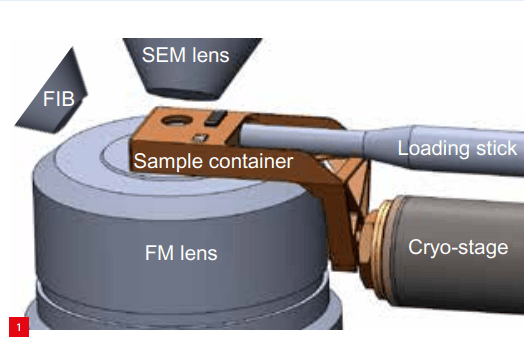
Delmic, located in Delft (NL), developed a fluorescence microscope (FM) that can be integrated with a scanning electron microscope (SEM). If the SEM is additionally provided with a focused ion beam (FIB) for preparing samples before imaging, the resulting system comprises three ‘lenses’ centred around the sample (Figure 1).